 |
|
Case Report
| ||||||
| Retroperitoneal schwannoma of the sciatic nerve: Case report and diagnostic review | ||||||
| Alberto Bouzón1, Ángela Iglesias2, Manuel Gómez1, Ángel Álvarez3, Juan Mosquera4 | ||||||
|
1Department of Surgery, San Rafael Hospital, A Coruña, Spain.
2Department of Radiology, San Rafael Hospital, A Coruña, Spain. 3Department of Plastic Surgery, San Rafael Hospital, A Coruña, Spain. 4Department of Anatomic Pathology, San Rafael Hospital, A Coruña, Spain. | ||||||
| ||||||
|
[HTML Abstract]
[PDF Full Text]
[Print This Article]
[Similar article in Pumed] [Similar article in Google Scholar] |
| How to cite this article: |
| Bouzón A, Iglesias Á, Gómez M, Álvarez Á, Mosquera J. Retroperitoneal schwannoma of the sciatic nerve: Case report and diagnostic review. J Case Rep Images Pathol 2016;2:25–28. |
|
Abstract
|
|
Introduction:
Schwannomas are usually benign tumors arising from Schwann cells of peripheral nerve sheaths. They are rarely located in the retroperitoneum and usually incidentally diagnosed. Sciatic nerve involvement is unusual.
Case Report: We report a case of schwannoma of the right sciatic nerve in a 39-year-old female with an incidental diagnosis. Magnetic resonance imaging (MRI) scan showed a 4.5 cm sciatic nerve dependent lesion located in the right pelvic region. Surgical complete resection of the lesion was performed. Final histopathological exam revealed the tumor to be a schwannoma. Conclusion: Schwannoma of the sciatic nerve can cause a pain in the lower limb similar to chronic sciatica. Magnetic resonance imaging (MRI) scan is helpful in the differential diagnosis. Histopathological examination gives definitive diagnosis. | |
|
Keywords:
Schwann cells, Schwannoma, Sciatic nerve
| |
|
Introduction
| ||||||
|
Schwannomas are rare tumors arising from Schwann cells of peripheral nerve sheaths. They are usually slow-growing benign tumors and rarely undergo malignant transformation. They occur most often in head, neck, posterior mediastinum and limbs. Retroperitoneal schwannomas account for about 2% of all retroperitoneal tumors [1] [2]. An accurate diagnosis at presentation is important to exclude a malignant retroperitoneal tumor. | ||||||
|
Case Report
| ||||||
|
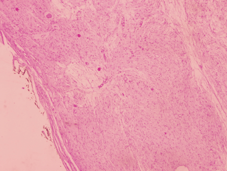
We present a case of 39-year-old female with a past medical history of chronic sciatica associated to a herniated disc. A right pelvic mass of 4.5 cm was incidentally diagnosed by ultrasound examination during a gynecological check. Computed tomography (CT) scan of the pelvis reported the presence of a 4.5 cm nodular lesion, located deep in the right pelvic region (Figure 1). Magnetic resonance imaging (MRI) scan showed a dependent right sciatic nerve lesion (Figure 2). The lesion contacted with the anterior surface of the sacrum and medially displaced the right hypogastric artery (Figure 3). A laparotomy to remove the lesion was scheduled. An encapsulated whitish retroperitoneal tumor attached to right sciatic nerve and easily resectable by blunt maneuvers was identified intraoperatively (Figure 4). The gross specimen was a smooth nodular lesion of 5x4 cm with soft consistency. The greyish cut surface showed the presence of cystic areas. Microscopic examination revealed a well-encapsulated lesion formed by spindle cells with eosinophilic cytoplasm and elongated nuclei without cytologic atypia or pleomorphism arranged in interlacing fascicles (Figure 5) and (Figure 6). Immunohistochemical staining showed diffuse positivity of tumor cells for S-100 protein, but was negative for smooth muscle actin (SMA). The patient had pain on the inside of the right foot during the immediate postoperative period. A demyelinating sensory neuropathy of the right posterior tibial nerve was diagnosed by electroneurogram. | ||||||
| ||||||
|
| ||||||
| ||||||
| ||||||
| ||||||
| ||||||
|
Discussion
| ||||||
|
Most retroperitoneal schwannomas are benign, and usually occur in patients between 20 and 50 years [3]. Some studies have reported a higher incidence in women [4] [5]. Sciatic nerve involvement is rare. Pelvic schwannomas are usually diagnosed incidentally, as solitary lesions, although some patients report abdominal pain or chronic sciatica. In case of chronic sciatica with no signs of radicular compression at MRI, the sciatic nerve must be radiologically examined all along its course [6]. Preoperative imaging tests are useful in determining the size, location and relationship of the lesion with neighboring tissues. Computed tomograhpy (CT) scan usually reveals a well-circumscribed lesion, with round or oval slight enhancement. Schwannomas are seen as isointense lesions on T1-weighted MRI and hyperintense on T2-weighted MRI scan. However, many schwannomas may show mix intensity on both T1- and T2-weighted images if they are associated with cystic degeneration, necrosis and (or) hemorrhage within the tumor [7]. Neurofibromas should be considered in the differential diagnosis. An eccentric association with the nerve is suggestive of a schwannoma. The definitive diagnosis of schwannoma is based on histopathologic and immunohistochemical findings [8]. Histologically, they are characterized by alternating areas of high and low cellularity, termed Antoni A and B regions. Mitotic figures are rarely observed. Large tumors often show cystic degeneration. Immunohistochemistry is positive for S-100, neuron specific enolasa and vimentin, but negative for SMA. Schwannomas are easily distinguished from leiomyomas or malignant peripheral nerve sheath tumors (NPNSTs). Tumor cells in leiomyomas are negative for S-100, but positive for SMA. NPNSTs have poorly differentiated cells with marked nuclear atypia and frequent mitoses. Surgical treatment represents the only option in symptomatic patients. The management of tumor involves surgical removal by refusing fascicular groups without penetrating them and preserving the continuity of the nerve [9]. Despite the complex location of most of the retroperitoneal tumors, the laparoscopic approach is increasingly used in the management of benign pelvic schwannomas [10]. Schwannomas have a good prognosis and low incidence of recurrence if the removal has been completed. | ||||||
|
Conclusion
| ||||||
|
Schwannomas of the sciatic nerve can cause pain in the lower limb similar to chronic sciatica. Although these tumors do not have specific imaging characteristics, radiologic examinations are helpful for achieving a diagnosis. Definitive diagnosis is possible only by histopathologic evaluation after surgical resection of the tumor. | ||||||
|
References
| ||||||
| ||||||
|
[HTML Abstract]
[PDF Full Text]
|
|
Author Contributions
Alberto Bouzón – Substantial contributions to conception and design, Acquisition of data, Analysis and interpretation of data, Drafting the article, Revising it critically for important intellectual content, Final approval of the version to be published Ángela Iglesias – Analysis and interpretation of data, Revising it critically for important intellectual content, Final approval of the version to be published Manuel Gómez – Analysis and interpretation of data, Revising it critically for important intellectual content, Final approval of the version to be published Ángel Álvarez – Analysis and interpretation of data, Revising it critically for important intellectual content, Final approval of the version to be published Juan Mosquera – Analysis and interpretation of data, Revising it critically for important intellectual content, Final approval of the version to be published |
|
Guarantor of submission
The corresponding author is the guarantor of submission. |
|
Source of support
None |
|
Conflict of interest
Authors declare no conflict of interest. |
|
Copyright
© 2016 Alberto Bouzón et al. This article is distributed under the terms of Creative Commons Attribution License which permits unrestricted use, distribution and reproduction in any medium provided the original author(s) and original publisher are properly credited. Please see the copyright policy on the journal website for more information. |
|
|


