 |
|
Case Report
| |||||
| Malignant pleural mesothelioma with metastases in the abdomen and left buttock: A case report | |||||
| Irene Moysset1, Gracia Valderas2, Ferran Losa3 | |||||
|
1MD, Department of Pathology, Consorci Sanitari Integral, L’Hospitalet de Llobregat, Barcelona, Spain
2MD, Department of Radiology, Consorci Sanitari Integral, L’Hospitalet de Llobregat, Barcelona, Spain 3MD, PhD, Department of Oncology, Consorci Sanitari Integral, L’Hospitalet de Llobregat, Barcelona, Spain | |||||
| |||||
|
[HTML Abstract]
[PDF Full Text]
[Print This Article] [Similar article in Pumed] [Similar article in Google Scholar] |
| How to cite this article |
| Moysset I, Valderas G, Losa F. Malignant pleural mesothelioma with metastases in the abdomen and left buttock: A case report. J Case Rep Images Pathol 2017;3:13–16. |
|
ABSTRACT
|
|
Introduction:
Malignant mesothelioma is a rare, aggressive neoplasia that affects the serous membranes. The pleural cavity is the most frequently affected area. Metastasis at the onset of the disease is rare, and muscular involvement is even more uncommon.
| |
| Keywords: Extrathoracic metastasis, Metastasis in skeletal muscle, Pleural mesothelioma | |
|
INTRODUCTION
| ||||||
|
Malignant mesothelioma is a rare neoplasia of mesodermal origin that affects the serous membranes of the pleura, peritoneum, pericardium, and tunica vaginalis, the most frequent location being the pleura [1]. It is commonly caused by exposure to asbestos [2][3]. Presentation with metastasis is rare as it appears more frequently during the clinical course of the disease, the most common sites being the lymph nodes, lungs, liver, adrenal glands, and kidneys [4]. At the subcutaneous and muscular level, metastasis is very rare; the majority of cases are due to local invasion [2]. In post-mortem studies, 87.7% of cases showed spreading of the disease [1]. | ||||||
|
CASE REPORT
| ||||||
|
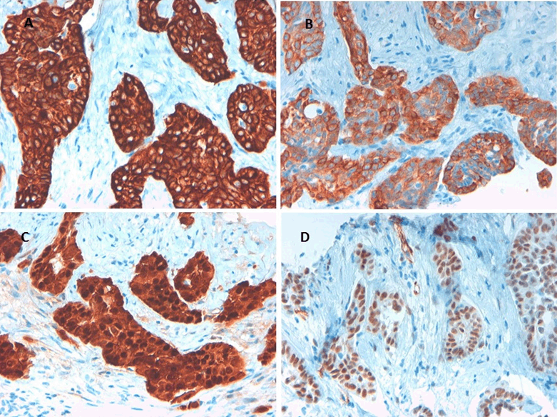
We report a case of a 64-year-old non-smoking male, who had had intermittent exposure to asbestos for three years. He had been undergoing treatment for hypertension. He presented with dyspnea on moderate exertion that had developed over several days, discomfort in the left hemithorax, and some intolerance to decubitus positions. The patient had a weight loss 10 kg in the last three months. A physical examination showed that he was conscious and lucid with normal coloring. Respiratory auscultation revealed hypophonesis in the lower half of the left hemithorax and right basal hemithorax. Hematological and hepatic findings were within the normal range. Tumor markers carbohydrate antigen (CA) 125, carcinoembryonic antigen (CEA), alpha-fetoprotein (AFP), CA 15-3, and CA 19-9 were shown to be within the normal range. Simple radiography of the posterior-anterior thorax showed bilateral pleural effusion with multiple nodular images in both hemithoraces (Figure 1). A contrast computed tomography (CT) scan of the thorax and abdomen revealed multiple bilateral pulmonary and pleural lesions, mediastinal lymphadenopathies (Figure 2), intra-abdominal implants (Figure 3), and a 56×40 mm polylobulated mass in the left buttock (Figure 4). A biopsy of the mass in the left buttock was made with an 18-gauge needle. Pathological study involved obtaining three cores of tissue measuring between 0.7 and 1 cm in length that were fixed in formalin. In the histological sections, we observed connective tissue infiltrated by epithelioid cells arranged in nests and trabeculae that showed atypia with hyperchromatic nuclei and prominent nucleoli. Occasional non-atypical mitosis was also detected (Figure 5). Immunohistochemical study revealed cytoplasmic positivity for keratins 5/6 and 7. Nuclear and cytoplasmic positivity was observed for calretinin, while nuclear positivity was detected for Wilms tumor 1 protein (Figure 6). Negative results were found for epithelial membrane antigen (Ber-Ep4), thyroid transcription factor-1 (TTF-1), protein suppressor p63, keratin 20, CEA, prostate-specific antigen (PSA), renal cell carcinoma (RCC), vimentin, common lymphoblastic leukemia antigen (CD10), S-100 protein, and CA 19.9. These results confirmed the diagnosis of metastasis of epithelioid mesothelioma. The patient passed away eight days later. | ||||||
| ||||||
| ||||||
|
DISCUSSION
| ||||||
|
Malignant mesothelioma is a rare and aggressive tumor. Most cases originate in the pleura. The most common age of presentation is between 50 and 70 years, and the presentation of the disease is most commonly localized. Clinical symptoms include dyspnea and non-pleural chest pain. Histologically, affected cells have been described as epithelioid, biphasic, and sarcomatoid, the former being the most frequent [5]. Presentation with distant metastasis of malignant pleural mesothelioma is rare, although some studies have demonstrated a 15% prevalence of extrathoracic metastasis using CT scans and positron emission tomography (PET) [6]. Metastasis in striated muscle involving solid tumors is very rare. The most frequent sites of metastasis are, in decreasing order, pulmonary, gastrointestinal, urological, genital, and mammary [7]. Six cases of malignant pleural mesothelioma with distant metastasis in striated muscle have been reported, three of which were detected at the onset of the disease [8][9][10][11][12][13]. The most frequent histological type in those cases was epithelioid. We report on a new case with distant extrathoracic metastasis, which is notable because of its unusual presentation. The rare site of metastasis in striated muscular tissue required a differential diagnosis among metastases of carcinoma, mesothelioma, and sarcoma. The definitive diagnosis was made based on histological and immunohistochemical study of the tumor located in the buttock. Furthermore, the clinical and radiological findings supported primary pleural mesothelioma. | ||||||
|
CONCLUSION
| ||||||
|
The presentation of malignant pleural mesothelioma with extrathoracic metastasis at disease onset is very rare, and our case was even more unusual as it involved metastasis in the skeletal muscle. Three cases have been previously reported. The type of presentation in our patient as well as the histological study required a differential diagnosis among metastases of carcinoma, mesothelioma, and sarcoma. The definitive diagnosis was based on microscopic and immunohistochemical study of the lesion in the buttock, which was important in selecting the correct treatment. | ||||||
|
REFERENCES
| ||||||
| ||||||
|
[HTML Abstract]
[PDF Full Text]
|
|
Author Contributions
Irene Moysset – Substantial contributions to conception and design, Acquisition of data, Analysis and interpretation of data, Drafting the article, Revising it critically for important intellectual content, Final approval of the version to be published Gracia Valderas – Analysis and interpretation of data, Revising it critically for important intellectual content, Final approval of the version to be published Ferran Losa – Acquisition of data, Revising it critically for important intellectual content, Final approval of the version to be published |
|
Guarantor of submission
The corresponding author is the guarantor of submission. |
|
Source of support
None |
|
Conflict of interest
Authors declare no conflict of interest. |
|
Copyright
© 2017 Irene Moysset et al. This article is distributed under the terms of Creative Commons Attribution License which permits unrestricted use, distribution and reproduction in any medium provided the original author(s) and original publisher are properly credited. Please see the copyright policy on the journal website for more information. |
|
|


