|
| Case Report | ||||||
| Metastatic breast cancer with GATA3, CK7 and ER positivity? A diagnostic pitfall | ||||||
| Diane-Ngan Trang1, Hongbo Wang2, Marlo Nicolas3, Alia Nazarullah1 | ||||||
| 1Department of Pathology and Laboratory Medicine, University of Texas Health, 7703 Floyd Curl Drive, MC#7750, San Antonio, Texas, USA 2Department of Pathology, Baylor Scott & White Medical Center, 2401 S. 31st Street, MS-01-266, Temple, Texas, USA 3Robert J. Tomsich Pathology and Laboratory Medicine Institute, Cleveland Clinic, Cleveland, Ohio, USA | ||||||
| ||||||
| [HTML Abstract]
[PDF Full Text][Print This Article] [Similar articles in PubMed][Similar articles in Google Scholar] |
| How to cite this article |
| Trang DN, Wang H, Nicolas M, Nazarullah. Metastatic breast cancer with GATA3, CK7 and ER positivity? A diagnostic pitfall. J Case Rep Images Pathol 2018;4:100021Z11DT2018. |
| ABSTRACT | ||||||
|
Introduction: Carcinoma of unknown primary can often be a challenge for clinicians and pathologists, and immunohistochemical stains (IHC) are often used as a useful diagnostic aid to narrow down the primary site of origin. In this context, co-expression of CK7, GATA3 and ER is highly suggestive of metastatic carcinoma from breast primary. This case highlights a major diagnostic pitfall in the interpretation of these stains. Case Report: A woman in her fifth decade presented with a possible thoracic vertebral lesion. The core biopsy showed cortical bone and a detached tissue with polygonal cells with eosinophilic cytoplasm and atypical,hyperchromatic nuclei with mild pleomorphism. These cells were strongly positive for CK7, GATA3, patchy positive for ER, and negative for CDX2, CK20, TTF1, thyroglobulin, mammaglobin, Melanin A, and PAX8 IHC stains. The interpretation based on these findings was metastatic breast adenocarcinoma. The patient received tamoxifen for stage 4 ER-positive breast cancer, in spite of no clinical or radiologic findings of a breast lesion. A 16 months after the initial diagnosis, the patient was referred to our institution. We noted prominent blood vessels and a few compressed glands positive for PAX8 resembling inactive endometrial glands within the detached fragment containing the atypical cells. The fact that intermediate and cytotrophoblasts express GATA3, cytokeratins and ER, along with the presence of inactive endometrial-type glands led us to conclude that the detached tissue likely represents a contaminant from an endometrial implantation site. Conclusion: This case reminds us that the immunohistochemical profile of trophoblasts is very similar to breast, and can cause major diagnostic pitfalls leading to erroneous diagnosis of metastatic breast cancer with severe consequences. IHC stains are not always specific, and should always be interpreted in the context of morphology. Keywords: Floater, GATA-3, Immunohistochemistry, Pitfall | ||||||
| INTRODUCTION | ||||||
|
“Floater,” an extraneous tissue fragment seen on a slide, are a common challenge encountered by surgical pathologists. In the majority of cases this can be easily resolved on the basis of the different histological findings of the intended section. However, challenges can arise when the floater is of the same tissue type or appears to be malignant. To aide in the resolution, pathologists often employ the use of immunohistochemical stains. GATA3 is a zinc-finger transcription factor is a highly sensitive marker for urothelial and breast carcinomas, but the specificity is relatively low [1],[2]. GATA3 is used predominantly to support the diagnosis of breast or urothelial origins in a carcinoma of unknown primary. GATA3 expression has been observed in 48–94% of breast carcinomas [1],[2],[3],[4]. In particular, the co-expression of CK7, GATA3, and ER is highly suggestive of breast cancer. However, GATA3 is also important for trophoblast differentiation [2], [4],[5],[6]. A study by Miettinenet al, demonstrates that trophoblastic tissue frequently expresses GATA3 [4]. The recognition that trophoblastic tissue also stains with GATA3 is important when dealing with suspected metastatic breast carcinomas. This case highlights a major diagnostic pitfall in the interpretation of these stains. | ||||||
| CASE REPORT | ||||||
|
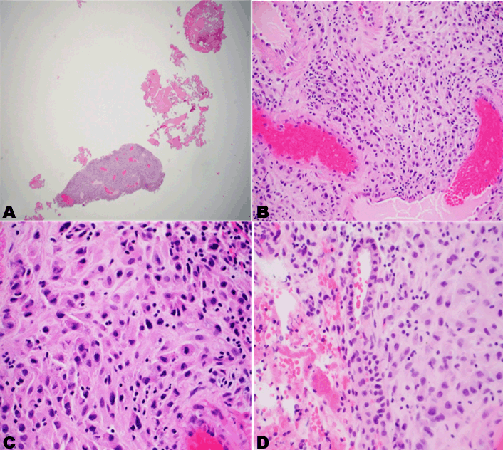
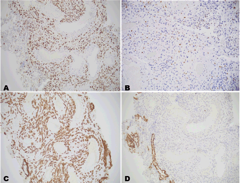
A woman in her 5th decade presented with a possible thoracic vertebral lesion following a compression fracture. A core biopsy of the lesion was performed. H&E slides and a panel of immunohistochemical stained slides were reviewed and sent to a reference lab for consultation. It was diagnosed as metastatic breast adenocarcinoma at both institutions. Although whole body imaging and PET scans failed to identify a primary tumor, the patient received hormonal therapy (Tamoxifen) for stage 4 ER-positive breast cancer. Over a year later, we received the slides for review Microscopic examination revealed dense bone and a detached fragment of tissue with predominantly plump polygonal cells, with abundant eosinophilic cytoplasm, and variably hyperchromatic atypical nuclei with mild nuclear pleomorphism. Prominent thick-walled blood vessels were noted amidst these polygonal cells. The periphery of this tissue showed few compressed glandular spaces Figure 1(A–D). The following immunohistochemical stains were performed: GATA3, ER, CK7, PAX-8, CDX2, CK20, TTF1, thryoglobulin, mammoglobin, Melanin A. Immunohistochemical stains show that the polygonal cells are strongly positive for CK7, GATA3, patchy positive for ER, and negative for CDX2, CK20, TTF1, thyroglobulin, mammaglobin, Melanin A, and PAX8. The epithelial cells of the compressed glandular component are strongly positive for PAX8, consistent with inactive endometrial glands Figure 2(A–D). Given the overall morphology and immunophenotype, the polygonal cells were determined to be most consistent with intermediate trophoblasts and cytotrophoblasts. There were nonreactive for CDX2, CK20, TTF1, thryoglobulin, mammoglobin, Melanin A. Based on histopathological and immunohistchemical findings, we concluded that the specimen represents a floater from a product of conception (POC)/implantation site. We performed a limited IHC panel on a known POC specimen and demonstrated the same immunoprofile (Figure 3). | ||||||
| ||||||
| ||||||
| ||||||
DISCUSSION | ||||||
|
Approximately 10% of all cancer patients develop spinal metastasis. Among the adult population 60% of spinal metastasis are of breast, lung, or prostate primaries [7] [8] [9]. When our patient was found to have a compression fracture with an initial diagnosis of metastatic breast adenocarcinoma, whole body imaging and PET scans were performed. Although a primary lesion was not identified, she received hormonal therapy for stage 4 ER positive breast cancer. In a study done by Gephardt and Zarbo, they reviewed 275 laboratories and found the frequency of exogenous tissue to be between 0.6% and 2.9% [10].Of those slides with extraneous tissue 25.3–59.5% of them had extraneous tissue located near diagnostic tissue [10]. Review of the literature does not reveal data reporting the frequency of extraneous tissue causing a misdiagnosis. Not all extraneous tissue will significantly impact the patient. Clues to help identify floaters of similar histological tissue type including location; such as being on a different plane of the main tissue or being along the edge of a slide. Furthermore, if the tissue is only on a single level it suggests a water bath contamination. When morphology is unhelpful, special methods such as DNA analysis and other methods described by Ritter et al. and Shibata may be helpful [11], [12]. Gephardt and Zarbo recommended cleaning the water baths before and during tissue section cutting and careful cleaning of the grossing station are paramount in limiting the number of floaters [10]. Cytokeratins 7 and 20 immunohistochemical staining profile is commonly used for broad differential diagnoses in the work up of carcinoma of unknown primary. A CK7+/CK20-immunoprofile is usually seen in tumors of lung, breast, endometrial, ovarian, thyroid, salivary gland, and upper gastrointestinal and pancreatobiliary primaries. Estrogen receptor-alpha is expressed in majority of breast and endometrial carcinomas, and less often in urothelial carcinomas. GATA3 is a sensitive and relatively specific marker for breast carcinoma. In one study done by Liu et al, GATA 3 expression was identified in 94% of invasive breast carcinoma [3] . GATA 3 expression is also seen in urothelial carcinomas in up to 86% cases as Liu et al reports [3]. Other GATA 3 positive tumors include choriocarcinoma, malignant mesothelioma, paraganglioma, pheochromocytoma, and salivary gland neoplasms [4], [13], [14]. An adenocarcinoma with co-expression of CK7, ER and GATA3 is highly specific for breast primary; however, rarely, urothelial carcinoma, choriocarcinoma, nephrogenic adenoma and sebaceous adenocarcinoma may show co-expression of these three markers. Immunohistochemistry is not useful for differentiating between normal trophoblastic tissue and breast carcinoma. There is overlap in their patterns of immunoreactivity. Both breast carcinomas and trophoblastic tissue are typically positive for CK7, ER, and GATA3. Other markers in this specific case that might be useful include: CD146, HLA-G and HSD3B1, which are all trophoblastic tissue specific markers [4], [5], [6], [13], [14]. | ||||||
| CONCLUSION | ||||||
|
It is important to recognize that the immunoprofile of GATA3, ER, CK7 positivity can be seen in breast neoplasms as well as trophoblasts in products of conception. Interpretation of immunohistochemistry should always be with caution, and in the context of morphology. | ||||||
| REFERENCES | ||||||
| ||||||
|
[HTML Abstract]
[PDF Full Text]
|
| Author Contributions
Diane-Ngan Trang – Substantial contributions to conception and design, Drafting the article, Critical revision of the article Hongbo Wang – Acquisition of data, Analysis and interpretation of data, Drafting the article Marlo Nicolas – Conception and design, Acquisition of data, Analysis and interpretation of data Alia Nazarullah – Conception and design, Acquisition of data, Analysis and interpretation of data, Drafting the article, Critical revision of the article, Final approval of the version to be published |
|
Guarantor of Submission
The corresponding author is the guarantor of submission. |
|
Source of Support
None |
|
Consent Statement
Written informed consent was obtained from the patient for publication of this case report. |
|
Conflict of Interest
Author declares no conflict of interest. |
|
Copyright
© 2018 Diane-Ngan Trang et al. This article is distributed under the terms of Creative Commons Attribution License which permits unrestricted use, distribution and reproduction in any medium provided the original author(s) and original publisher are properly credited. Please see the copyright policy on the journal website for more information. |
|
|