 |
Case Report
Solitary nodule of cutaneous reticulohistiocytosis: A case report
1 MD, MSc, Department of Pathology and Molecular Medicine, McMaster University, Hamilton, Ontario L8S 4K1, Canada
2 MD, FRCPC, FCAP, Assistant Professor, Department of Pathology and Molecular Medicine, McMaster University, Hamilton, Ontario L8S 4K1, Canada
3 MD, PhD, MSc, FRCPC, Associate Professor, Department of Pathology and Molecular Medicine, McMaster University, Hamilton, Ontario L8S 4K1, Canada
4 MB-Bch, FRCP, FCAP, FASCP, Professor, Department of Pathology and Molecular Medicine, McMaster University, Hamilton, Ontario L8S 4K1, Canada
Address correspondence to:
Jeffrey E Fournier
1280 Main Street West, Hamilton, Ontario L8S 4K1,
Canada
Message to Corresponding Author
Article ID: 100066Z11JF2022
Access full text article on other devices

Access PDF of article on other devices


How to cite this article
Fournier JE, Shao T, Popovic S, Alowami S. Solitary nodule of cutaneous reticulohistiocytosis: A case report. J Case Rep Images Pathol 2022;8(2):17–21.ABSTRACT
Introduction: Solitary cutaneous reticulohistiocytosis represents a rare form of benign monocyte/macrophage proliferation. On routine histology, these lesions are typically described as large cells with cytoplasm showing ground glass appearance and giant cells. They grow up to 1 cm in size with rare cases exceeding this size.
Case Report: This case report of a 28-year-old male demonstrated a nodule of reticulohistiocytosis measuring 2.2 cm in size. Microscopic features showed a well-demarcated nodule in the dermis with large histiocytes with ground-glass eosinophilic cytoplasm, giant cells, and foamy macrophages in a background of mixed inflammatory cells. Immunohistochemical staining showed positive staining for vimentin, CD68, CD31, with focal and patchy positivity for S100, CD43, and CD45 and negative staining for CD1a, langerin, CD21, CD23, CD30, CD34, ERG, D2-40, AE1/AE3, epithelial membrane antigen (EMA), smooth muscle actin (SMA), myogenin, desmin, SOX10, HMB-45, tyrosinase, and MelanA.
Conclusion: The microscopic and immunohistochemical findings are characteristic of this entity but it is important to recognize for proper management and differentiation from other malignant lesions.
Keywords: Cutaneous, Dermal, Reticulohistiocytosis, Solitary
Introduction
Histiocytoses refer to a group of rare disorders that include a proliferation and accumulation of monocyte/macrophage or dendritic cells [1]. Specifically, reticulohistiocytosis (previously referred to as reticulohistiocytoma) refers to solitary and multicentric histiocytic proliferations that have common histological findings of cells with abundant granular eosinophilic cytoplasm (ground glass appearance) mixed with giant cells, with or without systemic involvement respectively [2],[3]. Multiple case reports and few case series have been published on this entity, and it has been shown that the majority measure less than 1 cm with rare cases growing to larger sizes [4],[5],[6],[7]. This case report demonstrates microscopic and immunohistochemical findings of a 2.2 cm solitary cutaneous reticulohistiocytosis.
Case Report
A 28-year-old male patient presented to his family physician with a raised lesion in the left anterior axillary line in August 2021. The lesion was first noticed only months prior in May 2021 and had continued to grow and was reported to intermittently bleed. At the time of presentation, the lesion, a soft and raised nodule, measured 2.2×1.7 cm. No other lesions were identified or noted on clinical examination at the time of resection. When the lesion was resected, the clinical impression was that of a squamous cell carcinoma versus keratoacanthoma.
Microscopic examination demonstrated skin with a well-demarcated nodule involving the dermis and superficial subcutaneous tissue (Figure 1). It was mainly composed of a dense population of large histiocytes with glassy abundant, eosinophilic cytoplasm admixed with occasional giant cells and foamy macrophages (Figure 2). Many of the larger cells demonstrated emperipolesis throughout the lesion. This was in the background of mixed inflammatory cells including many eosinophils and neutrophils. The overlying epithelium appeared to be intact and uninvolved. There was minimal hemosiderin pigmentation consistent with prior hemorrhage.
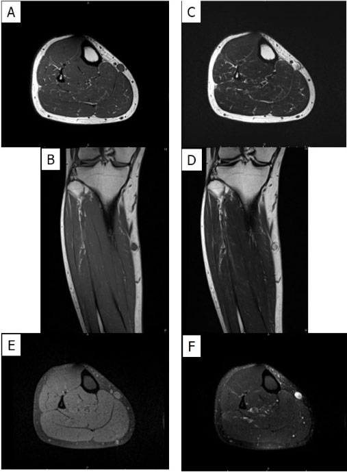
Immunohistochemical staining was performed and demonstrated positive staining for vimentin, CD68, and CD31 (Figure 3), with focal and patchy positivity for S100, CD43, and CD45 (Figure 4); and negative staining for CD1a, langerin, CD21, CD23, CD30, CD34, ERG, D2-40, AE1/AE3 (Figure 5), EMA, SMA, myogenin, desmin, SOX10, HMB-45, tyrosinase, and MelanA (Figure 6). Molecular studies were performed, including BRAF (exons 11 and 15), Kit (exons 8, 9, 10, 11, 13, 14, 17, and 18), KRAS (exons 2–4), and NRAS 2–4, and the lesion showed no clinically significant sequence variants detected in these regions.
A final diagnosis of “cutaneous histiocytic proliferation, favor reticulohistiocytosis” was rendered. The lesion was completely excised at the time of diagnosis and no further management or treatment was performed.


Discussion
Reticulohistiocytosis is a rare disorder of histiocytes with characteristic findings and three clinical presentations: solitary, diffuse, and multicentric [8]. These most commonly present clinically as multicentric with systemic involvement in the literature [9],[10],[11],[12]. Currently, the etiology of solitary cutaneous reticulohistiocytosis remains unclear, but it is believed to represent a reactive process to trauma, inflammatory process, or other unknown causes [2],[13],[14]. The differential diagnosis of these lesions includes other disorders such as Langerhans cell histiocytosis, Rosai–Dorfman disease, juvenile xanthogranuloma, necrobiotic xanthogranuloma, melanocytic lesions, and histiocytic sarcoma [1],[4].
Typically, these lesions present as a yellow to reddish-brown papule or nodule with a smooth surface lacking ulceration [13],[15]. Although the microscopic findings of “ground glass” eosinophilic cytoplasm and giant cells are characteristic, the differential histologically remains quite broad. It is therefore important to utilize immunohistochemical studies to differentiate from other benign and malignant conditions or conditions with genetic implications [16]. Regardless of the type, all forms of reticulohistiocytosis demonstrate the same pattern of immunohistochemical staining [2]. Typical immunohistochemical staining patterns show positivity for CD68, CD163, CD45, vimentin, and negativity for CD1a, langerin, and focal to negative S100 staining [4],[8],[17],[18].
Solitary cutaneous reticulohistiocytosis typically does not exceed 1 cm in diameter; however, rarely larger lesions have been reported [4],[19]. Diagnostic difficulties might arise clinically and histologically as a lesion larger than 1 cm may not be considered likely based on size alone. This pitfall may result in increased pathology reporting turnaround time, increased costs associated with immunohistochemical and other special ancillary tests, and increased patient anxiety while awaiting results of a biopsy or resection.
Once a diagnosis of reticulohistiocytosis is made, a full clinical examination and investigations should be undertaken to work up a patient for systemic involvement or other conditions [2]. As there are three clinical types of reticulohistiocytosis, a pathologist should not sub-classify based on a single biopsy or resection specimen. In such cases, a broad diagnosis of “reticulohistiocytosis” should be made. Solitary reticulohistiocytoses have been known to resolve on their own but may require surgical excision for symptomatic or cosmetic reasons [20],[21],[22]. Local recurrence is uncommon after incomplete surgical excision and is especially uncommon after complete surgical resection [4],[23].
Conclusion
This represents a case report of a large reticulohistiocytosis with characteristic microscopic and immunohistochemical findings. Although these lesions are benign and rare, with larger lesions being even rarer, it is important to produce a proper diagnosis for patients to receive appropriate management and to limit confusion with other malignant entities.
REFERENCES
1.
Luder CM, Nordmann TM, Ramelyte E, et al. Histiocytosis–Cutaneous manifestations of hematopoietic neoplasm and non-neoplastic histiocytic proliferations. J Eur Acad Dermatol Venereol 2018;32(6):926–34. [CrossRef]
[Pubmed]

2.
Bonometti A, Berti E; for Associazione Italiana Ricerca Istiocitosi ONLUS. Reticulohistiocytoses: A revision of the full spectrum. J Eur Acad Dermatol Venereol 2020;34(8):1684–94. [CrossRef]
[Pubmed]
3.
Cohen PR, Lee RA. Adult-onset reticulohistiocytoma presenting as a solitary asymptomatic red knee nodule: Report and review of clinical presentations and immunohistochemistry staining features of reticulohistiocytosis. Dermatol Online J 2014;20(3):doj_21725.
[Pubmed]
4.
Miettinen M, Fetsch JF. Reticulohistiocytoma (solitary epithelioid histiocytoma): A clinicopathologic and immunohistochemical study of 44 cases. Am J Surg Pathol 2006;30(4):521–8. [CrossRef]
[Pubmed]
5.
Purvis WE 3rd, Helwig EB. Reticulohistiocytic granuloma (reticulohistiocytoma) of the skin. Am J Clin Pathol 1954;24(9):1005–15. [CrossRef]
[Pubmed]
6.
Zelger B, Cerio R, Soyer HP, Misch K, Orchard G, Wilson-Jones E. Reticulohistiocytoma and multicentric reticulohistiocytosis. Histopathologic and immunophenotypic distinct entities. Am J Dermatopathol 1994;16(6):577–84. [CrossRef]
[Pubmed]
7.
Yamamoto T. Skin manifestation associated with multicentric reticulohistiocytosis. J Clin Rheumatol 2022;28(1):e234–9. [CrossRef]
[Pubmed]
8.
Shibuya R, Tanizaki H, Kaku Y, et al. A plaque-type solitary reticulohistiocytoma in a two-year-old boy. Case Rep Dermatol 2015;7(1):7–9. [CrossRef]
[Pubmed]
9.
Heathcote JG, Guenther LC, Wallace AC. Multicentric reticulohistiocytosis: A report of a case and a review of the pathology. Pathology 1985;17(4):601–8. [CrossRef]
[Pubmed]
10.
Farabi B, Jamgochian M, Rao BK. Multicentric reticulohistiocytosis with dermatomyositis-like features: A case report with dermoscopy and reflectance confocal microscopy findings. J Cutan Pathol 2022;49(4):388–92. [CrossRef]
[Pubmed]
11.
Sanchez-Alvarez C, Sandhu AS, Crowson CS, et al. Multicentric reticulohistiocytosis: The Mayo Clinic experience (1980–2017). Rheumatology (Oxford) 2020;59(8):1898–905. [CrossRef]
[Pubmed]
12.
Bhuvitha MS, Nandakumar G. Multicentric reticulohistiocytosis – A rare clinicopathological entity. J Evolution Med Dent Sci 2021;10(33):2854–8.
13.
de Oliveira FL, Nogueira LLC, Chaves GMC, et al. A unique dermoscopy pattern of solitary cutaneous reticulohistiocytosis. Case Rep Dermatol Med 2013;2013:674896. [CrossRef]
[Pubmed]
14.
Luz FB, Kurizky PS, Ramos-e-Silva M. Reticulohistiocytosis. Dermatol Clin 2007;25(4):625–32. [CrossRef]
[Pubmed]
15.
Delorenze LM, Russo T, Boechat M, et al. Dermoscopy of solitary cutaneous reticulohistiocytoma. G Ital Dermatol Venereol 2018;153(4):579–80. [CrossRef]
[Pubmed]
16.
Pallu IR, Proença PGL, de Souza Boscoli S, et al. Uncommon cutaneous solitary reticulohistiocytoma: A case report. Biomed J Sci & Tech Res 2021;37(4):29704–6. [CrossRef]
17.
Luz FB, Gaspar AP, Ramos-e-Silva M, et al. Immunohistochemical profile of multicentric reticulohistiocytosis. Skinmed 2005;4(2):71–7. [CrossRef]
[Pubmed]
18.
Trotta F, Castellino G, Monaco AL. Multicentric reticulohistiocytosis. Best Pract Res Clin Rheumatol 2004;18(5):759–72. [CrossRef]
[Pubmed]
19.
Ghosh SK, Bandyopadhyay D, Ghosh A, Bar C. Multiple yellowish plaques and nodules in a young man: A case of multiple cutaneous reticulohistiocytomas. J Turk Acad Dermatol 2009;3:93202c.
20.
Chisolm SS, Schulman JM, Fox LP. Adult xanthogranuloma, reticulohistiocytosis, and Rosai-Dorfman disease. Dermatol Clin 2015;33(3):465–73. [CrossRef]
[Pubmed]
21.
Kieselova K, Santiago F, Amado C, Henrique M. Case for diagnosis. Solitary violaceous nodule on the toe. An Bras Dermatol 2018;93(4):595–7. [CrossRef]
[Pubmed]
22.
Behera B, Kumari R, Thappa DM, Gochhait D. Dermoscopic features of a case of solitary reticulohistiocytoma. Indian J Dermatol Venereol Leprol 2020;86(4):435–8. [CrossRef]
[Pubmed]
23.
Mannan AASR, Kahvic M, Singh NG, Zahir M. Solitary epithelioid histiocytoma (reticulohistiocytoma) of the glans penis. Int Urol Nephrol 2012;44(5):1345–8. [CrossRef]
[Pubmed]
SUPPORTING INFORMATION
Author Contributions
Jeffrey E Fournier - Conception of the work, Design of the work, Drafting the work, Revising the work critically for important intellectual content, Final approval of the version to be published, Agree to be accountable for all aspects of the work in ensuring that questions related to the accuracy or integrity of any part of the work are appropriately investigated and resolved.
Tiffany Shao - Analysis of data, Revising the work critically for important intellectual content, Final approval of the version to be published, Agree to be accountable for all aspects of the work in ensuring that questions related to the accuracy or integrity of any part of the work are appropriately investigated and resolved.
Snezana Popovic - Analysis of data, Revising the work critically for important intellectual content, Final approval of the version to be published, Agree to be accountable for all aspects of the work in ensuring that questions related to the accuracy or integrity of any part of the work are appropriately investigated and resolved.
Salem Alowami - Conception of the work, Design of the work, Revising the work critically for important intellectual content, Final approval of the version to be published, Agree to be accountable for all aspects of the work in ensuring that questions related to the accuracy or integrity of any part of the work are appropriately investigated and resolved.
Guarantor of SubmissionThe corresponding author is the guarantor of submission.
Source of SupportNone
Consent StatementWritten informed consent was obtained from the patient for publication of this article.
Data AvailabilityAll relevant data are within the paper and its Supporting Information files.
Conflict of InterestAuthors declare no conflict of interest.
Copyright© 2022 Jeffrey E Fournier et al. This article is distributed under the terms of Creative Commons Attribution License which permits unrestricted use, distribution and reproduction in any medium provided the original author(s) and original publisher are properly credited. Please see the copyright policy on the journal website for more information.


