 |
Case Report
Cylindroma with extensive stromal adipocytic metaplasia likely arising in nevus lipomatosus superficialis: A rare entity
1 Department of Pathology and Molecular Medicine, McMaster University, Hamilton, Ontario, Canada
2 Libyan International Medical University, Benghazi, Libya
3 Division of Plastic Surgery, McMaster University, Hamilton, Ontario, Canada
4 Department of Pathology and Molecular Medicine, St. Joseph’s Healthcare Hamilton, McMaster University, Hamilton, Ontario, Canada
Address correspondence to:
Andrea Chen
Department of Pathology and Molecular Medicine, McMaster University, HSC-2N10, 1280 Main Street West, Hamilton, ON L8S 4K1,
Canada
Message to Corresponding Author
Article ID: 100098Z11AC2026
Access full text article on other devices

Access PDF of article on other devices


How to cite this article
Chen A, Alowami M, Boutross-Tadross O, Cin AD, Alowami S. Cylindroma with extensive stromal adipocytic metaplasia likely arising in nevus lipomatosus superficialis: A rare entity. J Case Rep Images Pathol 2026;12(1):6–9.ABSTRACT
Introduction: Cylindromas are uncommon benign dermal adnexal tumors that tend to arise in the scalp of middle-aged females. Nevus lipomatosus superficialis is a rare hamartomatous connective tissue nevus consisting of mature adipocytes in the reticular dermis.
Case Report: Herein, we report a case of a 53-year-old woman with a congenital scalp lesion demonstrating histopathological features consistent with a benign cylindroma arising within a nevus lipomatosus superficialis.
Conclusion: This case highlights the diagnostic challenge associated with recognizing nevus lipomatosus superficialis in conjunction with other dermal tumors. This is the second reported case of a cylindroma arising from nevus lipomatosus superficialis, in what otherwise would be considered a cylindroma with associated extensive stromal adipocytic metaplasia. The patient’s unique presentation expands the understanding and association of cylindromas with nevus lipomatosus superficialis. Complete surgical excision of the lesion is considered curative treatment, as both entities are benign.
Keywords: Adipocytic metaplasia, Benign adnexal tumors, Cylindroma, Nevus lipomatosus superficialis
Introduction
Nevus lipomatosus superficialis (NLS), also known as nevus lipomatosus cutaneous superficialis (NLCS), or a pedunculated lipofibroma, is a rare hamartomatous connective tissue nevus consisting of mature adipocytes in the reticular dermis. It was first described by Hoffmann and Zurhelle in 1921 [1]. These lesions can be present from birth or infancy, and although pathogenesis and etiology is not fully understood, it has been theorized to develop from possible perivascular origin of young, immature adipocytes into mature fat, akin to fetal adipogenesis, as compared to previous presumed local ectopic or heterotopic development of mature adipocytic tissue [2],[3]. Two variants of NLS are typically characterized clinically; classic and solitary. Classic NLS is described as multiple grouped skin colored papular polypoid dermal based lesions, most often developing congenitally or within the first two decades of life, mostly in the posterior surface pelvic girdle regions, including lower back, buttocks, and upper thighs [3],[4]. The solitary variant primarily develops as single papular lesion in adults, and can appear anywhere on the skin, similarly found on the posterior surface of the body, on the back, buttocks, and upper thighs, as well as the axilla and scalp [4].
Nevus lipomatosus superficialis has been associated with other dermal lesions such as folliculosebaceous cystic hamartomas, angiokeratomas, and dermoid cysts along with other pilar abnormalities, however, rarely are associated with dermal appendageal tumors [3],[5].
Cylindromas are uncommon benign dermal adnexal tumors that tend to arise in the scalp of middle-aged females. Yu and Alowami previously reported the first case of cylindroma arising within a nevus lipomatosus superficialis [6]. Herein, we present the second reported case of a benign cylindroma arising from a nevus lipomatosus superficialis.
Case Report
A 53-year-old woman presented with a small mass on her scalp since birth, described as previously only slightly bothering her, only recently becoming bothersome from a functional (tenderness) aspect. Her past medical history included diabetes and hypertension. Physical examination revealed a skin colored mass on the vertex of the scalp, with multiple small vessels. There was no significant erythema, and no swollen or inflamed areas. No purulence or fluctuance was appreciated, and there was no pain on palpation. Excisional biopsy was performed.
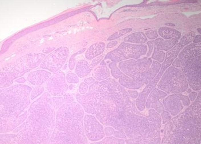
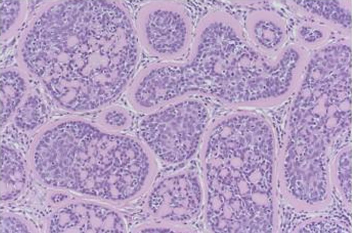
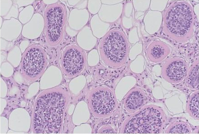
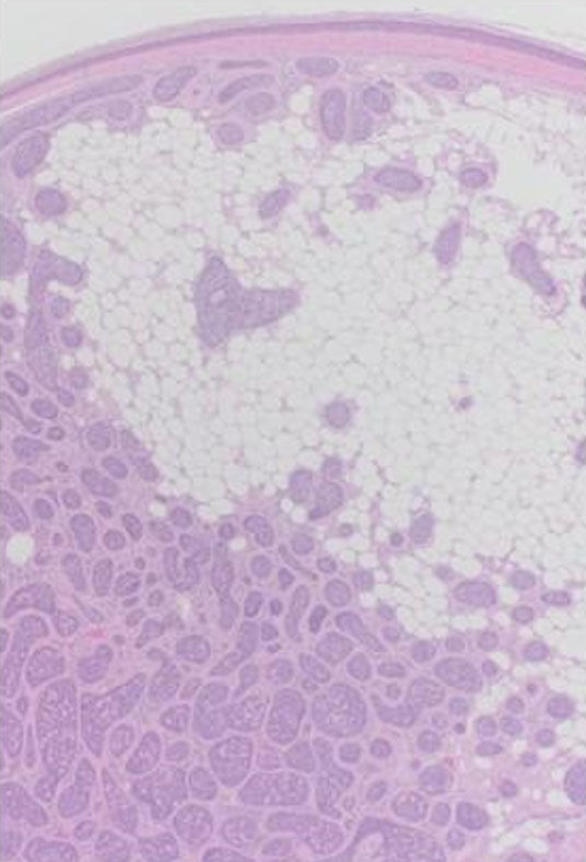
Microscopic examination showed a poorly circumscribed, unencapsulated dermal based tumor composed of multiple variably sized, irregularly shaped basaloid nests and islands of basaloid cells, separated and ensheathed by an eosinophilic hyalinized band like basement membrane, forming the characteristic “Jigsaw puzzle” like pattern of a cylindroma (Figure 1 and Figure 2). A band of compressed papillary dermis was appreciated separating the dermal based basaloid islands from the overlying epidermis, without any appreciable connection. The islands were composed of smaller, bland appearing basaloid cells with hyperchromatic nuclei mostly located at the periphery of the islands, with larger paler cells with vesicular shaped nuclei within the central areas. Focal duct like formations were seen, along with globules of brightly eosinophilic hyaline basement material within the islands. Near the periphery of the tumor, mature adipocytes were seen separating the dermal basaloid nests with surrounding eosinophilic bands, expanding the stroma between nests, distributed circumferentially up towards the papillary dermis (Figure 3 and Figure 4). No necrosis, nuclear atypia, or mitotic figures were identified.
Histomorphological findings were consistent with a benign cylindroma with extensive adipocytic metaplasia versus arising from within a nevus lipomatosus superficialis.
Discussion
The current case can be compared to the one other previously described cylindroma arising from nevus lipomatosus superficialis, as reported by Yu and Alowami [6]. The cases presented similarly in women within the fifth and sixth decades of life. Both lesions were located on the scalp. Our case presented with a solitary skin colored papular lesion on the scalp from a young age, largely asymptomatic until pain and tenderness resulted in clinical presentation and excision. This could be indicative of a congenital, longstanding solitary NLS lesion with a cylindroma arising or developing within the lesion.
It is postulated that this entity only being previously described once could be possibly due to the diagnostic challenge in recognizing the nevus lipomatosus superficialis component with mature adipocytes within the reticular dermis. The cylindroma element is distinct and recognizable; however in such cases, differentiating a cylindroma arising from a nevus lipomatosus superficialis compared to what is presumed to be a cylindroma with associated stroma adipocytic or lipomatous metaplasia can be difficult. Focal mature adipocytic fat stromal metaplasia or deposition can be attributed to degenerative or reactive changes in connective tissue, however in our case, the extensive and circumferential pattern of the mature adipocytes in the stromal tissue, almost encircling the cylindroma nests make it more likely to be arising in a previous nevus lipomatosus superficialis, similar to the previously reported case [6].
This contrasts to nevus sebaceus (nevus of Jadassohn) and other follicular hamartomas, which are hamartomatous lesions that present on the scalp in infancy, with abnormalities of the folliculo-sebaceous glands. Nevus sebaceus has been commonly associated with benign adnexal tumors such as trichoblastomas and trichoepitheliomas, syringocystadenoma papilliferum, as well as uncommonly basal cell carcinoma and other malignant transformations [7],[8]. No similar associations with adnexal tumors have been made with NLS; however, perhaps in reporting this second known case of a cylindroma associated and arising in NLS, this may lead to increased recognition and understanding of the pathogenesis and associations of an otherwise rare entity.
As both entities are considered benign (NLS and cylindroma), complete surgical excision is considered curative treatment, with recurrences extremely rare and often only in incomplete excisions.
Conclusion
This case highlights the diagnostic challenge associated with recognizing nevus lipomatosus superficialis, especially in the context of other dermal tumors, as in this case of cylindroma. This is the second reported case of a cylindroma arising from nevus lipomatosus superficialis, in what otherwise would be considered a cylindroma with associated extensive stromal adipocytic metaplasia. The patient’s unique presentation, including age and lesion, expands the understanding and association of cylindromas with nevus lipomatosus superficialis.
REFERENCES
1.
Hoffmann E, Zurhelle E. Über einen Naevus lipomatodes cutaneus superficialis der linken Glutäalgegend. Archiv für Dermatologie und Syphilis 1921;130(1):327–33. [CrossRef]

2.
Reymond JL, Stoebner P, Amblard P. Nevus lipomatosus cutaneous superficialis. An electron microscopic study of four cases. J Cutan Pathol 1980;7(5):295–301. [CrossRef]
[Pubmed]
3.
Goucha S, Khaled A, Zéglaoui F, Rammeh S, Zermani R, Fazaa B. Nevus lipomatosus cutaneous superficialis: Report of eight cases. Dermatol Ther (Heidelb) 2011;1(2):25–30. [CrossRef]
[Pubmed]
4.
Lima CDS, Issa MCA, de Souza MB, Góes HFO, Santos TBPD, Vilar EAG. Nevus lipomatosus cutaneous superficialis. An Bras Dermatol 2017;92(5):711–3. [CrossRef]
[Pubmed]
5.
Bancalari E, Martínez-Sánchez D, Tardío JC. Nevus lipomatosus superficialis with a folliculosebaceous component: Report of 2 cases. Patholog Res Int 2011;2011:105973. [CrossRef]
[Pubmed]
6.
Yu R, Alowami S. Cylindroma with stromal adipose tissue metaplasia versus arising in a background of nevus lipomatosus. Case Rep Pathol 2014;2014:203298. [CrossRef]
[Pubmed]
7.
Cribier B, Scrivener Y, Grosshans E. Tumors arising in nevus sebaceus: A study of 596 cases. J Am Acad Dermatol 2000;42(2 Pt 1):263–8. [CrossRef]
[Pubmed]
8.
Kamyab-Hesari K, Seirafi H, Jahan S, Aghazadeh N, Hejazi P, Azizpour A, et al. Nevus sebaceus: A clinicopathological study of 168 cases and review of the literature. Int J Dermatol 2016;55(2):193–200. [CrossRef]
[Pubmed]
SUPPORTING INFORMATION
Acknowledgments
No generative AI technology has been used in this paper.
Author ContributionsAndrea Chen - Conception of the work, Design of the work, Acquisition of data, Drafting the work, Final approval of the version to be published, Agree to be accountable for all aspects of the work in ensuring that questions related to the accuracy or integrity of any part of the work are appropriately investigated and resolved.
Moaz Alowami - Conception of the work, Design of the work, Drafting the work, Final approval of the version to be published, Agree to be accountable for all aspects of the work in ensuring that questions related to the accuracy or integrity of any part of the work are appropriately investigated and resolved.
Odette Boutross-Tadross - Conception of the work, Design of the work, Revising the work critically for important intellectual content, Final approval of the version to be published, Agree to be accountable for all aspects of the work in ensuring that questions related to the accuracy or integrity of any part of the work are appropriately investigated and resolved.
Arianna Dal Cin - Drafting the work, Final approval of the version to be published, Agree to be accountable for all aspects of the work in ensuring that questions related to the accuracy or integrity of any part of the work are appropriately investigated and resolved.
Salem Alowami - Conception of the work, Design of the work, Acquisition of data, Revising the work critically for important intellectual content, Final approval of the version to be published, Agree to be accountable for all aspects of the work in ensuring that questions related to the accuracy or integrity of any part of the work are appropriately investigated and resolved.
Guarantor of SubmissionThe corresponding author is the guarantor of submission.
Source of SupportNone
Consent StatementWritten informed consent was obtained from the patient for publication of this article.
Data AvailabilityAll relevant data are within the paper and its Supporting Information files.
Conflict of InterestAuthors declare no conflict of interest.
Copyright© 2026 Andrea Chen et al. This article is distributed under the terms of Creative Commons Attribution License which permits unrestricted use, distribution and reproduction in any medium provided the original author(s) and original publisher are properly credited. Please see the copyright policy on the journal website for more information.

