 |
Case Report
Perianal hidradenoma papilliferum in a male: A case report and review of the literature
1 Medical Student, Department of Surgery, Bendigo Health, 100 Barnard Street, Bendigo, Victoria 3550, Australia
2 Pathology Registrar, Anatomical Pathology, Australian Clinical Labs, 1868 Dandenong Road, Clayton, Victoria 3168, Australia
3 General Surgery Registrar, Department of Surgery, Bendigo Health, 100 Barnard Street, Bendigo, Victoria 3550, Australia
4 Pathology Consultant, Anatomical Pathology, Australian Clinical Labs, 100 Barnard Street, Bendigo, Victoria 3550, Australia
Address correspondence to:
Lily Li
Department of Surgery, Bendigo Health, 100 Barnard Street, Bendigo, Victoria,
Australia
Message to Corresponding Author
Article ID: 100101Z11LL2026
Access full text article on other devices

Access PDF of article on other devices


How to cite this article
Li L, Wong HS, Taylor D, Fernando R. Perianal hidradenoma papilliferum in a male: A case report and review of the literature. J Case Rep Images Pathol 2026;12(1):22–26.ABSTRACT
Introduction: Hidradenoma papilliferum is a rare, benign, predominantly cystic tumor that predominantly affects women between 30 and 60 years of age. It is thought to originate from anogenital mammary-like glands, and therefore most commonly arises in the anogenital region. Clinically, it typically presents as a slow-growing, solitary, circumscribed nodule measuring between 0.5 cm and 2.0 cm in diameter.
Case Report: We report a rare case of a perianal hidradenoma papilliferum in a 70-year-old male who presented with a 2 cm highly pedunculated perianal lesion at the anal verge. This lesion had reportedly been present for several months and had recently increased in size. A literature review was completed and a total of 15 cases of hidradenoma papilliferum were identified in men, of which only three involved the perianal region. The majority of cases (9/15) were located in the head and neck region.
Conclusion: We therefore report a new case of this rare entity. To our knowledge, this is only the fourth documented case of perianal hidradenoma papilliferum in a male patient.
Keywords: Benign tumor, Hidradenoma papilliferum, Papillary hidradenoma, Perianal
Introduction
Hidradenoma papilliferum (HP) is a rare, benign cystic tumor that occurs predominantly in women aged 30 to 60 [1]. It is thought to originate from anogenital mammary-like glands, and therefore most commonly arises in the anogenital region with vulvar lesions reported approximately four times more frequently than perianal lesions [2]. Rare ectopic cases have also been reported, typically in the head and neck. Interestingly, while anogenital HP occurs almost exclusively in women, ectopic forms demonstrate a more balanced sex distribution, with nearly half of reported cases arising in men [3]. Here, we report a rare case of perianal HP in a 70-year-old man and review the literature on male HP. To our knowledge, this is only the fourth documented case of perianal HP in a male patient.
Case Report
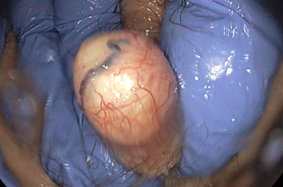
A 70-year-old man presented to our General Surgery clinic for a surveillance colonoscopy following previous findings of multiple tubular adenomas with low-grade dysplasia. During the procedure, a 2 cm polyp at the anal verge was incidentally identified but not removed (Figure 1). On return to clinic for discussion of the colonoscopy results, further assessment of the anal lesion was undertaken.
On examination, there was a highly pedunculated mass measuring approximately 2 cm in diameter at the 11 o’clock position. This lesion had reportedly been present for several months and had been recently increasing in size. There was no history of bleeding, pain, or pruritus. The patient’s past medical history included obesity, hypertension, and hypercholesterolemia.
Pathology Findings with IHC Confirmation
Surgical excision under general anesthesia was performed for histological evaluation. Informed consent was obtained from the patient for publication of this case report and accompanying images.
Gross Findings
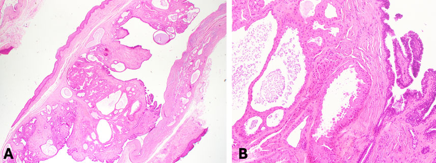
Soft tissue specimen measuring 18 × 12 × 4 mm. The specimen was sectioned into five pieces for histological examination.
Histopathological Findings
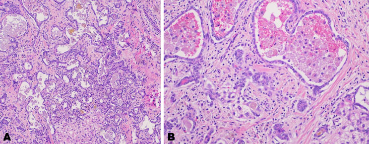
Microscopic examination demonstrated a polypoid lesion with anal squamous epithelial covering (Figure 2). There was a well demarcated cystic lesion in the dermis lined by double layered epithelium with myoepithelial cells and inner cuboidal to columnar cells with apocrine snouts. Focally, there was an intramural solid area with tubular and cystic structures with benign eccrine and apocrine epithelium. Low papillary-like structures were also seen. There were no features of atypia or malignancy.
Immunohistochemistry (IHC) Findings
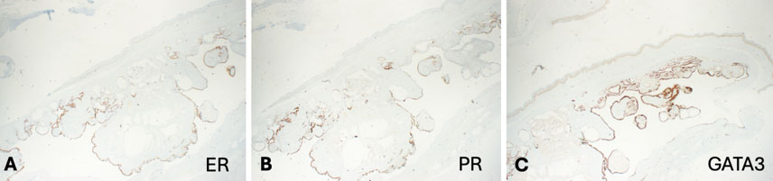
Immunohistochemistry showed tumor cells positive for GATA3, estrogen receptor, and progesterone receptor (Figure 3). Taken together, the morphological and immunohistochemical findings supported a diagnosis of HP.
Discussion
Though first described in 1878, HP remains under-recognized, and its pathogenesis poorly understood. Typically presenting as an asymptomatic, slow-growing, raised nodule, some patients may report pain, pruritus, ulceration, and bleeding [2]. Lesions usually range from 0.5 cm to 2.5 cm in diameter, though sizes up to 10 cm have been reported [4].
Initially believed to arise from apocrine sweat glands, current evidence suggests that HP originates from anogenital mammary-like glands which are morphologically similar to breast tissue but are normally present in the interlabial sulcus [2],[5]. Like normal breast tissue, most lesions express estrogen and progesterone receptors and are influenced by female sex hormones, likely accounting for their predominance in post-pubertal women. Nevertheless, cases have also been documented in men and at ectopic sites. Ectopic HP is believed to arise from modified apocrine glands such as the glands of Moll at the eyelid margin or the ceruminous glands of the external auditory canal [6],[7]. While HP in women is strongly linked to the anogenital region, cases in men are more frequently ectopic. Additionally, the role of sex hormones in the pathogenesis of male HP is thought to be less significant than in women; however, our patient’s tumor did demonstrate positivity for both estrogen and progesterone receptors. More research is required to clarify the contribution of hormonal pathways to HP development in both sexes.
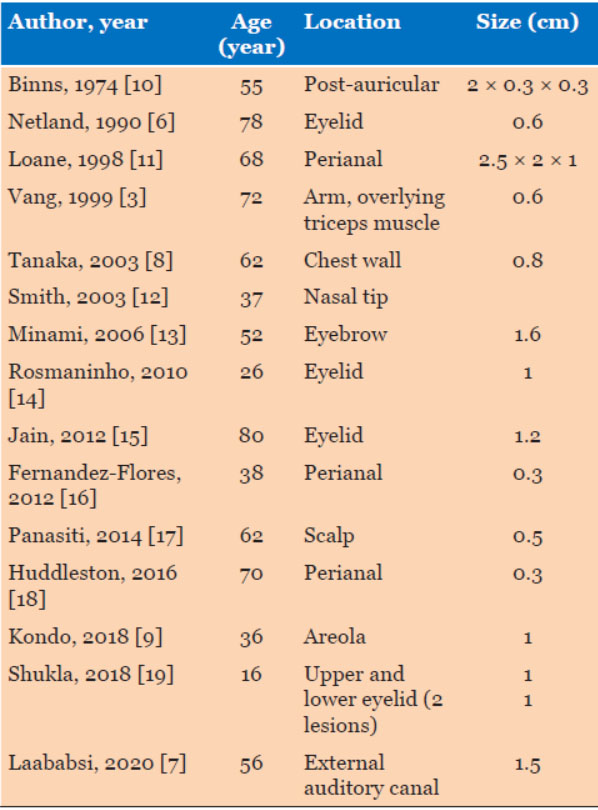
To better characterise HP in the male population, we conducted a review of the literature and identified 15 case reports of HP in men (Table 1). Only three of these cases involved the perianal region and thus, our case represents the fourth reported instance, highlighting the rarity of the condition.
Consistent with the literature, the majority of ectopic HP in our review occurred in the head and neck region (9/15, 60%) though other sites such as the arm, chest wall, and areola were also described [3],[8],[9]. We found that in men, perianal HP seemed to present at a higher mean age compared with ectopic HP (61.5 years vs 52.5 years). This observation differs slightly from existing literature, which reports that ectopic HP presents on average 10–20 years later than anogenital lesions [3]. Considering our exclusive focus on male patients and the limited number of reported cases, these findings should be interpreted with caution. Nevertheless, clinicians should be aware of the possibility of HP in an older male presenting with a perianal lesion.
The overall prognosis of HP is excellent, with surgical excision being curative in the vast majority of cases. Rare instances of local recurrence have been described [20], and there are occasional reports of malignant transformation in the forms of mammary-type hidradenocarcinoma, adenocarcinoma in situ and adenosquamous carcinoma [21],[22],[23]. Human papillomavirus (HPV), particularly HPV-16, has been detected in some of these lesions; however, a causal relationship has not yet been established [24].
Conclusion
In conclusion, we describe a fourth case of male perianal HP, a rare benign tumor most commonly found in women. It is often asymptomatic and may be overlooked or misdiagnosed due to its lack of distinctive clinical features. Surgical excision remains necessary for both definitive diagnosis and curative treatment.
REFERENCES
1.
Daniel F, Mahmoudi A, de Parades V, Fléjou JF, Atienza P. An uncommon perianal nodule: Hidradenoma papilliferum. Gastroenterol Clin Biol 2007;31(2):166–8. [CrossRef]
[Pubmed]

2.
Hama M, Oiso N, Kawada A. Ulcerated hidradenoma papilliferum. Int J Dermatol 2013;52(2):198–9. [CrossRef]
[Pubmed]
3.
Vang R, Cohen PR. Ectopic hidradenoma papilliferum: A case report and review of the literature. J Am Acad Dermatol 1999;41(1):115–8. [CrossRef]
[Pubmed]
4.
Hernández-Angeles C, Nadal A, Castelo-Branco C. Hidradenoma papilliferum of the vulva in a postpartum woman: A case report. J Obstet Gynaecol 2017;37(5):683–4. [CrossRef]
[Pubmed]
5.
El-Khoury J, Renald MH, Plantier F, Avril MF, Moyal-Barracco M. Vulvar hidradenoma papilliferum (HP) is located on the sites of mammary-like anogenital glands (MLAGs): Analysis of the photographs of 52 tumors. J Am Acad Dermatol 2016;75(2):380–4. [CrossRef]
[Pubmed]
6.
Netland PA, Townsend DJ, Albert DM, Jakobiec FA. Hidradenoma papilliferum of the upper eyelid arising from the apocrine gland of Moll. Ophthalmology 1990;97(12):1593–8. [CrossRef]
[Pubmed]
7.
Laababsi R, Elkrimi Z, Bouzbouz A, Lekhbal A, Rouadi S, Abada R, et al. Hidradenoma papilliferum of the external auditory canal. Case report. Ann Med Surg (Lond) 2019;49:41–43. [CrossRef]
[Pubmed]
8.
Tanaka M, Shimizu S. Hidradenoma papilliferum occurring on the chest of a man. J Am Acad Dermatol 2003;48(2 Suppl):S20–1. [CrossRef]
[Pubmed]
9.
Kondo RN, Melhado IP, Moreira CR, Crespigio J. Ectopic hidradenoma papilliferum. An Bras Dermatol 2018;93(3):474–5. [CrossRef]
[Pubmed]
10.
Binns JH. A rare case of hidradenoma papilliferum: Report of a case and review of the literature. Br J Plast Surg 1974;27(4):367–9. [CrossRef]
[Pubmed]
11.
Loane J, Kealy WF, Mulcahy G. Perianal hidradenoma papilliferum occurring in a male: A case report. Ir J Med Sci 1998;167(1):26–7. [CrossRef]
[Pubmed]
12.
Smith FB, Shemen LJ, Guerrieri C, Ismail SS. Hidradenoma papilliferum of nasal skin. Arch Pathol Lab Med 2003;127(2):E86–8. [CrossRef]
[Pubmed]
13.
Minami S, Sadanobu N, Ito T, Natsuaki M, Yamanishi K. Non-anogenital (ectopic) hidradenoma papilliferum with sebaceous differentiation: A case report and review of reported cases. J Dermatol 2006;33(4):256–9. [CrossRef]
[Pubmed]
14.
Rosmaninho ADN, de Almeida MTDP, Costa V, Sanches MMV, Lopes C, Selores Gomes Meirinhos MM. Ectopic hidradenoma papilliferum. Dermatol Res Pract 2010;2010:709371. [CrossRef]
[Pubmed]
15.
Jain D, Siraj F, Grover AK, Garg KK. Hidradenoma papilliferum presenting as an eyelid mass. Ophthalmic Plast Reconstr Surg 2012;28(6):e152–3. [CrossRef]
[Pubmed]
16.
Fernandez-Flores A, Valerdiz S. Pseudocarcinomatous hyperplasia associated with hidradenoma papilliferum. Am J Dermatopathol 2012;34(3):e31–6. [CrossRef]
[Pubmed]
17.
Panasiti V, Curzio M, Roberti V, Lieto P, Gobbi S, Devirgiliis V, et al. Ectopic hidradenoma papilliferum dermoscopically mimicking a blue nevus: A case report and review of the literature. Int J Dermatol 2014;53(2):e103–6. [CrossRef]
[Pubmed]
18.
Huddleston MK, Jenkins CP, Nelson EC. Hidradenoma papilliferum: A case report of an uncommon perianal lesion. Am Surg 2016;82(2):E43–4.
[Pubmed]
19.
Shukla P, Malaviya AK. Ectopic hidradenoma papilliferum of eyelid: A rare entity with diagnostic challenge. Indian J Pathol Microbiol 2018;61(2):287–9. [CrossRef]
[Pubmed]
20.
Spindler L, Pommaret E, Moyal Barracco M, Fathallah N, Plantier F, Duchatelle V, et al. Anal and vulvar hidradenoma papilliferum are similar: A study of 14 cases. [Article in French]. Ann Dermatol Venereol 2019;146(8–9):537–41. [CrossRef]
[Pubmed]
21.
Kim GY, Solanki MH, Guo R. Vulvar apocrine hidradenocarcinoma arising in a hidradenoma papilliferum—A case report. J Cutan Pathol 2021;48(8):1085–7. [CrossRef]
[Pubmed]
22.
Shah SS, Adelson M, Mazur MT. Adenocarcinoma in situ arising in vulvar papillary hidradenoma: Report of 2 cases. Int J Gynecol Pathol 2008;27(3):453–6. [CrossRef]
[Pubmed]
23.
Bannatyne P, Elliott P, Russell P. Vulvar adenosquamous carcinoma arising in a hidradenoma papilliferum, with rapidly fatal outcome: Case report. Gynecol Oncol 1989;35(3):395–8. [CrossRef]
[Pubmed]
24.
Vazmitel M, Spagnolo DV, Nemcova J, Michal M, Kazakov DV. Hidradenoma papilliferum with a ductal carcinoma in situ component: Case report and review of the literature. Am J Dermatopathol 2008;30(4):392–4. [CrossRef]
[Pubmed]
SUPPORTING INFORMATION
Author Contributions
Lily Li - Conception of the work, Design of the work, Acquisition of data, Analysis of data, Drafting the work, Revising the work critically for important intellectual content, Final approval of the version to be published, Agree to be accountable for all aspects of the work in ensuring that questions related to the accuracy or integrity of any part of the work are appropriately investigated and resolved.
Hui Sze Wong - Conception of the work, Design of the work, Acquisition of data, Drafting the work, Revising the work critically for important intellectual content, Final approval of the version to be published, Agree to be accountable for all aspects of the work in ensuring that questions related to the accuracy or integrity of any part of the work are appropriately investigated and resolved.
Danielle Taylor - Conception of the work, Design of the work, Revising the work critically for important intellectual content, Final approval of the version to be published, Agree to be accountable for all aspects of the work in ensuring that questions related to the accuracy or integrity of any part of the work are appropriately investigated and resolved.
Ruchira Fernando - Acquisition of data, Analysis of data, Revising the work critically for important intellectual content, Final approval of the version to be published, Agree to be accountable for all aspects of the work in ensuring that questions related to the accuracy or integrity of any part of the work are appropriately investigated and resolved.
Guarantor of SubmissionThe corresponding author is the guarantor of submission.
Source of SupportNone
Consent StatementWritten informed consent was obtained from the patient for publication of this article.
Data AvailabilityAll relevant data are within the paper and its Supporting Information files.
Conflict of InterestAuthors declare no conflict of interest.
Copyright© 2026 Lily Li et al. This article is distributed under the terms of Creative Commons Attribution License which permits unrestricted use, distribution and reproduction in any medium provided the original author(s) and original publisher are properly credited. Please see the copyright policy on the journal website for more information.
